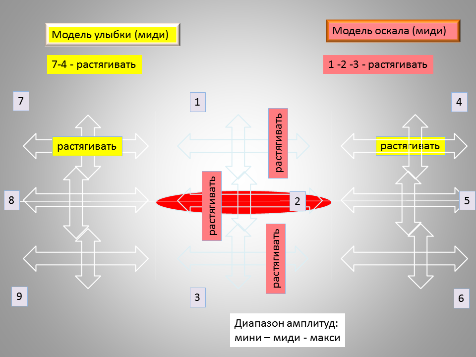
Да, требуется короткое внятное сенсорное описание, с указанием чётко наблюдаемых признаков. Признаки должны быть качественно порядка - они не должны отсылать к необходимости проведения каких-то измерений.
Ну при улыбке уголки губ вверх
"Вверх" - по сравнению с чем? В этом месте спряталось какое-то сравнение= измерение.
а при оскале на том же уровне что и обычно.
Это предполагает напряжение одинаковых лицевых мышц и при улыбке, и при оскале. Но, такое предположение неверно.
Точные определения экспрессия рта. Экспр. кайфа и "рег
metanymous в посте Metapractice (оригинал в ЖЖ)
Наблюдения за гориллами показали, что при игре...
Множество - десятки и сотни различных контекстов, в которых мы можем ожидать от горилл неких позитивных чувств. Но, из этого числа надо вычесть проективные/провоцирующие человеческие проективные эмоции.
В моём постинге я предложил рассматривать задачу, в первую очередь, с человеческой экспрессией.
они открывают пасть и прикрывают зубы руками, как бы говоря: «Я мог бы тебя укусить, но не буду».
Открывать пасть/рот можно десятками разных способов.
Совершать экспрессивные манипуляции возле рта руками можно сотней разных способов.
Такую гримасу учёные соотносят со смехом.
Спрашивается, какую "такую" конкретно гримасу учёные проективно соотносят с собственным смехом? Ибо, "смех" животных они и не думали описывать.
А иногда гориллы широко улыбаются, открывая оба ряда зубов почти до голливудского стандарта. Наблюдения показали, что такая «улыбка» появляется, когда обезьяна хочет попросить о чём-то другого или сгладить неловкость.
"Широко" - является параметром, требующим проведения измерения. Что же касается моделирования, то в нём наиболее ценные качественные параметры.
Если игра затягивается или становится слишком рискованной, горилла начинает «улыбаться» напарнику — и это служит дополнительным мирным сигналом, удерживающим шуточную потасовку от превращения в настоящую драку. Чем грубее и резче движения, чем активнее игра, тем чаще приходится сглаживать возникающие неловкости, тем чаще требуется подтверждение статуса и субординации и тем чаще на мордах у горилл появляются улыбки.
Именно так. Таким манером ученые "слепили" два недоказанных/не объективированных наблюдения в одно и получили вывод, что улыбка есть по происхождению агрессия. Или она "родственна" агрессии. И т.п.
Между тем, и гориллы по-своему, и люди по-своему совершенно по разному раскрывают свои пасти/рты в ситуациях/контекстах, в которых:
--центральным фактором являются и остаются во всех случаях ресурсные переживания. А ежели стимулы для ресурсных переживаний прекращаются, то и тот час же прекращается соответствующая экспрессия
--центральным фактором являются нересурсные переживания, например, необходимость контролировать малоресурсное/пороговое поведение сородичей. Экспрессивные сигналы для этого случая требуются радикально другими - совершенно отличными от экспрессии гипотетических "улыбок". В противном случае, такой контролирующей экспрессии всегда не будет достаточно и каждый раз только уже натурально агрессией можно будет тормозить зарвавшихся в некоей активности субъектов.

Следует определить: улыбается или же слегка скалится известный актёр?
Вопрос сводится к определению подобия/противоположности между улыбкой и оскалом.
Или, требуется описать визуально-экспрессивную модель улыбки, сравнив её с таковой в отношении оскала:
Улыбка
Улыбка - оскал

Армен Григорян
Первый, второй и третий человек
https://books.google.ru/books?id=Si2YCgAAQBAJ&pg=PA78&lpg=PA78&dq=%D1%81%D0%BC%D0%B5%D1%85*%D0%B0%D0%B3%D1%80%D0%B5%D1%81%D1%81%D0%B8%D1%8F*%D0%BA%D0%BE%D0%BD%D1%80%D0%B0%D0%B4+%D0%BB%D0%BE%D1%80%D0%B5%D0%BD%D1%86&source=bl&ots=QE9NvBAvlI&sig=7R7misRqbVOu4OfKDvFDl9aFaTc&hl=ru&sa=X&ved=0ahUKEwiZ3OXzm_DKAhVmApoKHSH6Dh4Q6AEIJjAC#v=onepage&q=%D1%81%D0%BC%D0%B5%D1%85*%D0%B0%D0%B3%D1%80%D0%B5%D1%81%D1%81%D0%B8%D1%8F*%D0%BA%D0%BE%D0%BD%D1%80%D0%B0%D0%B4%20%D0%BB%D0%BE%D1%80%D0%B5%D0%BD%D1%86&f=false
![[pic]](https://dl.dropboxusercontent.com/s/uv66zbhifj6htjo/%D0%A1%D0%BC%D0%B5%D1%85%20-%20%D0%B0%D0%B3%D1%80%D0%B5%D1%81%D1%81%D0%B8%D1%8F.png?dl=0)
СОУВЮР (Cмех - Оптимизм - Улыбка - Веселье - Юмор - Радость) (5) Смех vs агрессия
metanymous в Metapractice (оригинал в ЖЖ)
Смотрел краем глаза передачу, в которой целый час доказывали, что смех есть производная агрессии. Т.е. экспрессия смеха произошла от экспрессии агрессии. Сильно удивился. Но, потом посмотрел в гуугл: "смех*агрессия" -
https://www.google.ru/search?q=http://metapractice.livejournal.com/friends&ie=utf-8&oe=utf-8&rls=org.mozilla:ru:unofficial&client=seamonkey-a&gws_rd=cr&ei=yRq8Vp3MHsX76ATG87-oCg#newwindow=1&q=%D1%81%D0%BC%D0%B5%D1%85*%D0%B0%D0%B3%D1%80%D0%B5%D1%81%D1%81%D0%B8%D1%8F
Заметно, что в гуугле развёрнуто окно овертона "смех=агрессия". Начали, как положено в таких случаях, издалека. Типа, ещё Дарвин указывал на эволюционную связь смеха и агрессии.
Но, я всё равно не согласен. И в данном случае несогласие может быть выражено только через краткую формальную модель смеха. Вот, ежели удастся показать, что формальная модель смеха радикально отличается от агрессии, то наша взяла. В противном случае результат очевиден.


Оригинал взят у davydov_index в Люди, ставшие гениями после удара по голове

Большинство из нас мечтают проснуться в один прекрасный день и обнаружить у себя новый талант, умение или знание иностранного языка. Некоторым довелось пережить такое в реальности. Один из них – Джейсон Пэджетт.
Джейсон Пэджетт: математика и физика
Недоучившись в средней школе, Джейсон работал в мебельном магазине своего отца, а ночами пропадал на вечеринках. Пока в один прекрасный день его не ударили по голове и 31-летний молодой человек внезапно стал гением в физике и математике.
У Джейсона диагностировали синдром саванта (савантизм), при котором мозговые травмы могут внезапно подарить пострадавшему неожиданные таланты в математике, изобразительном искусстве или в музыке.
43-летний Пэджетт один из немногих людей в мире, способных от руки рисовать фракталы – самоподобные геометрические фигуры с громадным количеством повторяющихся звеньев. На создание одного фрактала Джейсону требуются недели, а то и месяцы.

Джейсон Пэджетт
Орландо Серрелл: календарные вычисления
Играя в бейсбол, 10-летний Орландо Серрелл был сбит с ног ударом мячика по голове. Мальчик не рассказал об этом родителям, и врачи его не осмотрели.
В течение целого года Орландо страдал от непрекращающихся головных болей. Когда же боли наконец прекратились, мальчик обнаружил удивительную способность: он мог абсолютно точно назвать день недели по дате.
Вообще, любые вычисления, связанные с календарем, Серрелл выполняет в мгновение ока. Он моментально может назвать количество дней между любыми двумя датами, или же, например, сколько раз в течение 1000 лет 12 марта выпадало на четверг.
Орландо Серрелл
Томми Макхью: поэзия и изобразительное искусство
Бывший заключенный, 51-летний Томми Макхью сумел выжить после двух субарахноидальных кровоизлияний мозга в 2001 году.
Повреждения в лобной и височной долях мозга заставили Томми постоянно рифмовать слова и писать картины. Когда закончились холсты, Макхью начал расписывать стены своего дома.
Я чувствую в себе какую-то женственность. Моя голова теперь полна рифм, картин и различных образов.
– Томми Макхью
Тони Чикория: музыка
Хирурга-ортопеда Тони Чикорию ударила молния в 1994 году во время прогулки по парку. Находящейся поблизости медсестре удалось спасти ему жизнь, и здоровье доктора Чикория постепенно восстановилось.
Вскоре врач обнаружил необъяснимое стремление слушать классические музыкальные произведения на фортепиано, а затем играть и самому. При этом, до тех пор Чикория никогда и близко не подходил к какому-либо музыкальному инструменту.
Он начал покупать ноты, самостоятельно научился играть на фортепиано, а затем принялся за сочинение сложных музыкальных пьес.
Тони Чикория
Алонзо Клемонс: скульптура
Еще в младенческом возрасте Алонзо Клемонс получил серьезную травму головы после падения на пол в ванной комнате.
Обладая IQ 40 и будучи неспособным читать и писать, он вырос в приюте для детей с ограниченными возможностями.
С раннего возраста он продемонстрировал удивительный талант к скульптуре. Стоило ему увидеть какое-нибудь животное хоть на несколько секунд, он тут же лепил его из подручных материалов, например, из мыла.
Теперь 56-летний Алонзо – известный американский скульптор, работы которого продаются за десятки тысяч долларов.
Алонзо Клемонс
Бен Макмахон: китайский язык
Когда Бен Макмахон попал в кому после автомобильной аварии, его родители опасались, что он может из нее никогда не выйти.
Однако уже через неделю австралийский студент пришел в себя – и заговорил на китайском, точнее на его севернокитайском диалекте (мандарин).
Все как в тумане, помню только, что когда я проснулся и увидел медсестру-китаянку, то сразу подумал, что нахожусь в Китае. Будто во сне, я вдруг заговорил по-китайски.
– Бен Макмахон, 22 года
До этого Бен проходил базовый курс китайского в школе, но не закончил его. В конце концов, ему вновь удалось вспомнить английские слова, но неожиданные способности к китайскому не исчезли.
В настоящее время Бен работает гидом по Австралии для китайских туристов и ведет телепередачу на китайском для иммигрантов.
Бен Макмахон
Лахлан Коннорс: музыкальный вундеркинд
У Лахлана Коннорса, к большому огорчению его матери, с детства начисто отсутствовал музыкальный слух. Даже сыграть собачий вальс на фортепиано ему не удавалось.
Зато подросток увлекался спортом, что и привело дважды к серьезным сотрясениям мозга во время игры в лакросс. После этого у Лахлана начались эпилептические припадки и галлюцинации, так что с контактным спортом пришлось распрощаться.
Однако мозговые травмы привели к неожиданному побочному эффекту: 17-летний Коннорс внезапно обнаружил способность играть на различных музыкальных инструментах, не прилагая к этому практически никаких усилий.
Специалисты считают, что у подростка, вероятно, наблюдаются эпилептические припадки, подобные тем, что на протяжении всей своей жизни испытывал великий композитор и виртуоз Фредерик Шопен.
Подросток, которому еще пару лет назад «медведь на ухо наступил», теперь играет на 13 различных музыкальных инструментах, включая фортепиано, гитару, укулеле и гармонику.
Лахлан Коннорс
Дэниел Тэммет: математика
Испытав в три года эпилептический припадок, Дэниел Тэммет помешался на числах.
В школе он выигрывал призы по математике, однако его необычные способности были обнаружены лишь в 25 лет. Специалисты диагностировали у Тэммета высокофункциональный аутизм с синдромом саванта, который позволял ему буквально жонглировать с числами.
Так, например, он правильно назвал 22 514 цифр в числе пи в течение 5 часов и 9 минут. Он также разговаривает на 10 языках, включая исландский, который Дэниел выучил на спор в течение недели.
Процесс вычислений в уме 35-летний Дэниел описывает следующим образом:
Когда я перемножаю два числа, то вижу их в форме двух картинок. Затем образ начинает изменяться и развиваться, и внезапно появляется третья. Это и есть ответ. Я вычисляю, практически не думая об этом, лишь представляя в уме картинки.
Дэниел Тэммет
naked-science
Он был стандартным полиглотом до травмы./…/
Благодаря Вилли мы, кстати, нашли общее в судьбах полиглотов. Во-первых, любовь к языкам у них рождается в детстве. Во-вторых, растут они, как правило, в многоязыковой семье или среде. То есть с ранних лет для них мир звучит на разных языках. И главное — цель, которую они себе ставят.
Дочитали до конца.


![[pic]](http://naked-science.ru/sites/default/files/images/maxresdefault_0.jpg)


